Alexandre Barros
CV:
Dr. Alexandre Barros. PhD Tisue Engineering, Regenerative Medicine and Stem Cells from University of Minho, Portugal, in collaboration with the Department of Bioengineering, University of California, Berkeley, United States of America; MSc in Technology and Properties of Polymers, at the University of Minho, Portugal and BSc in Biotechnology at the Polytechnic institute of Viana do Castelo, Portugal. During his Ph.D., Alexandre was granted with several awards has received the major innovation award in Portugal the Novo Banco Innovation Award. The research developed during the Ph.D. and post-doctoral period originate a spin-off company calls HydrUStent, that are pursuing the clinical trials now. Since April of 2018, Alexandre Barros is IP & Innovation Manager at I3Bs, 3B's Research Group, University of Minho, Portugal with an IP portfolio with more than 120 international patents. He has published 19 peer-reviewed articles, 35 communications in international conferences, receiving three times the award of Best Oral Presentation, 2 book chapter and is co-inventor of 10 international patent. He has google scholar h-index of 10 and received over 382 citations.
HydrUStent - From bench to business
HydrUStent was born in 2016 with the mission to create, develop and manufacture biodegradable medical devices and help to build a sustainable world.
Over the past couple of years, we have successfully developed innovative medical devices for urology and gastroenterology. Our patented technology allowed us to create the first 100% biodegradable hydrogel-based ureteral stent (HydrUStentTM). This product eliminates the need for a second surgery, avoids bacterial infection/encrustation and reduces treatment cost by 60%. Furthermore, we also provide medical device development services.
We provide engineering services for biodegradable and urological medical devices design and prototype development. Our team will help to develop your product at any step of the way, from early concept to preclinical prototypes.
Ali Abou-Hassan
Short biography: Dr. Ali Abou-Hassan is an Associate Professor at Sorbonne University, Paris, France. His research deals with strategies combining microfluidics and inorganic physical-chemistry. Fascinated by out of equilibrium phenomena he also studies the growth of materials in a biomimetic approach assisted by microfluidics and nanomaterial bio-modifications in cells using material science approaches.
Affiliation: Sorbonne Université, CNRS, Laboratoire Physicochimie des Electrolytes et Nanosystèmes InterfaciauX, UMR 8234, Campus Pierre et Marie Curie, 4 place Jussieu F-75005 Paris
Physico-chemical multi-scale microfluidic approach to kidney stones formation
Over the past decades, the increase in kidney stone (KS) formers has raised the importance to understand the biomineralization process responsible for urolithiasis. Calcium oxalate (CaOx) crystallization – KS main inorganic compound – has largely been characterized under batch synthesis conditions that cannot be regarded as biomimetic with respect to the microscale environment in the kidney and to the urinary flow. A chemical model considering these features would provide, in fine, the biomedical community with a predictive platform for suitable and reliable diagnosis.
In this work, we used a reversible microchannel to mimic the collecting duct in the nephron where CaOx stones can form due to supersaturated levels in calcium and oxalate ions. Within the channel, CaOx crystallization was induced under co-laminar mixing of Ca2+(aq) and Ox2-(aq) ions matching pathological concentrations – i.e. hypercalciuria and moderate hyperoxaluria. Scanning electron microscopy and Raman spectroscopy were used to support our investigations. They showed that CaOx crystals precipitate in a mixture of monohydrated whewellite (CaC2O4.H2O, COM) and dihydrated weddellite (CaC2O4.2H2O, COD) in the microchannel, similar to what is observed by the physicians. In situ information on the kinetics of CaOx crystal growth could be acquired in our microfluidic system. They confirmed the effect of the hydrodynamic conditions as well as of the presence of chelating molecules on the growth kinetics and the final chemistry (phase, shape) of the formed CaOx crystals. Finally, in a trial to achieve a more complex biomimetic model (formation of KS on a Randall’s plaque), hydroxyapatite was grown in the microchannel and the CaOx crystal formation was investigated.
References
(1) D. Bazin et al. Characterization and some physico-chemical aspects of pathological micro-calcifications,
Chem. Rev. 2012, 112, 5092 - 5120.
(2) A. Abou-Hassan et al., Angew. Chem. Intl. Ed. 2010, 49, 6268-6286.
(3) G. Laffite et al., Lab on Chip, 16, 1157 (2016).

Bastien Chopard
Bastien Chopard is full professor in the computer science department, University of Geneva, and member of the Swiss Institute of Bioinformatics. His main research interests are the modeling and simulation of complex systems, HPC and biomedical applications.
Biomedical applications with the Lattice Boltzmann method
This talk presents different numerical models describing complex biomedical flow processes, with their clinical relevance. These model are based on the Lattice Bolzmann method which will be quickly introduced. Then we will describe following applications: (1) the flow of red blood cells and the transport of platelets, as a function of various blood pathology; (2) thrombus formation in cerebral aneurysms induced by a flow diverter, (3) assessment of the success or failure of a thrombolysis treatment in case of a stroke event; (4) characterization of a vertebroplasty intervention.
Daniela Casoni
Short biography: Daniela Casoni, DVM, Dr.Med.Vet, Dip. ECVAA, PhD. Affiliation: Head of Experimental Surgery Facility- Department of Biomedical Research (DBMR)- Faculty of Medicine- University of Berne.
Research interests: comparative pain physiopathology, pain therapy and neuroanaesthesia
“In vivo” models for urology surgery: How can the animals help us? How can we help them?
Animals represent a very important learning source in the research world. This consideration applies to several disciplines, including urology. Nonetheless, the ethics of using animals in research is a very actual matter of debate. Researchers and animalists are often standing on very different positions that seem incompatible. It is possible to conciliate them? Using animals in research is definitely a privilege of which the researchers should be aware. Animals must be used with great care and consciousness. In animal research, a rigorous methodological approach is required, including selecting the appropriate species, address any confounder that may interfere with the outcome and reporting the result with transparency and completeness of information. Looking the research achievements form a different perspective, we could also think that not only humans but also other animals could benefit out of animal studies, as a model of pathologies or treatment for pathologies affecting several species. Urethral stents are a typical example of this possibility. Approaching the research with less “ego” and more “eco” could maybe be the way to reconcile researchers, animals and the public opinion.
Dario Carugo
Dario Carugo is a Lecturer at the University of Southampton, where he coordinates the Micro & Nano Therapies (MiNaTher) research group. He holds a visiting position at the University of Oxford (Institute of Biomedical Engineering), where he previously worked as a PostDoctoral researcher. His expertise is in bio-microfluidics, with application in interventional medicine and ultrasound-mediated therapies. He develops therapeutic devices and agents for the treatment of cancerous and non-cancerous disorders, including infectious diseases, vascular and urinary dysfunctions, and tissue injuries. In the field of endourology, he develops numerical and experimental models to investigate the mechanisms of stent encrustation and design innovative stent architectures. He has received awards from the Institution of Mechanical Engineers, Department of Health, and EPSRC.
Developing a pre-clinical pipeline to innovate endourological devices: from microfluidics to full-scale models
Urinary stents are clinically deployed to retrieve urine drainage in the presence of ureteric occlusions. However, stenting is often associated with complications caused by encrustation and biofilm formation, which can compromise a patient’s quality of life and significantly impact on healthcare costs. Despite novel materials and coatings have been introduced to increase stent’s lifetime, encrustation is still recognised as a major and recurrent cause of failure.
Current pre-clinical models often lack the ability to replicate the complex dynamics of the urological flow mechanics, or to allow investigation of the upstream/downstream effects of the deployment of a urological device. These limitations have hindered the usage of these models as a pre-clinical platform to innovate current urological devices.
In this talk we will propose a multi-scale methodological pipeline, encompassing both experimental and computational (CFD) methods, to investigate the role of urinary flow dynamics on the deposition and growth of crystalline and bacterial deposits in stents. We will discuss the utility of these methods in the context of pre-clinical urological stent development, and present avenues for future research.
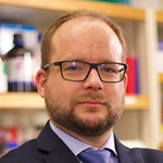
Dirk Lange
From Bench to Bedside: Preclinical Validation Using Large-Animal Models
The importance for in vivo validation of bench-top research results.
Specific large-animal models to test novel ureteral stent technologies.
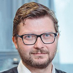
Dominik Obrist
ARTORG Center for Biomedical Engineering Research
University of Bern
Switzerland
Short bio:
Dominik Obrist does research on biomedical flow systems. He studied mechanical engineering at ETH Zurich (1991-1997) and earned a PhD in applied mathematics from the University of Washington. in Seattle (2000). In 2013, he was appointed Professor for Cardiovascular Engineering at the University of Bern.
Combining clinical data with computational models of biomedical flow systems
Dominik Obrist, ARTORG Center for Biomedical Engineering Research, University of Bern
Clinical data provide direct in vivo information on biomedical flow systems (e.g. the urinary tract). It may comprise information on flow rate and pressure from urodynamic testing or morphological information from imaging modalities. However, this data is typically noisy and incomplete (e.g. limited resolution of imaging modality) such that it cannot provide a complete picture of the whole system.
In contrast, computational models of biomedical flow systems (forward models) can very accurately model the underlying physical processes. At the same time, their clinical applicability is strongly limited by the lack of patient-specific data on morphology and physiological parameters.
We will discuss a concept for combining sparse and noisy clinical data with forward models, in order to create patient-specific simulations of biomedical flow systems. These data-driven predictions are the best possible approximation to the measured data, while fitting at the same time the underlying laws of physics of the forward model.
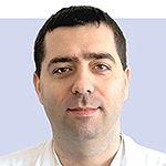
Duje Rako
Duje Rako MD, FEBU was promoted MD in 2006 at Zagreb University School of medicine and passed urology board exam in Zagreb, Croatia in 2013 and FEBU exam in Brussels in 2015. Currently working as consultant urological surgeon in private practice in Zagreb, Croatia and also occasionally at Countess of Chester NHS Foundation Trust in Chester, UK with previous experience at University hospital Dubrava in Zagreb, Croatia. Has special interest in urinary stones and endoscopy and is leader of workgroup State of art of urinary stents in ENIUS COST project.
Urologist’s perspective on urinary stents
This talk will focus on urologist’s perspective on urinary stents.
There are many situations urologists use stents in urinary system ranging from benign to malignant. Each situation caries unique challenges but also there are some common ones. We are going to discuss problems with stent placement and removal, stent irritation, encrustation, occlusion, migration just to name few. Urinary stone disease is most common cause for using urinary stents and its worldwide prevalence ranges from 1 to 20% with probable mean of 10%. This means that roughly one in ten of worldwide population could in some time in their life be candidate for urinary stent!!! It is therefore of paramount importance to focus on new solutions for old problems with urinary stents.
At the end of talk you should be able to recognise most of complications when using stents which should boost your interest in trying to replicate problem, identify its causes, research options and ultimately offer solution.
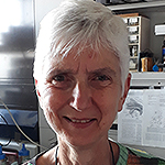
Els van Asselt
Short biography: Els van Asselt is a researcher at the Department of Urology of the Erasmus Medical Center in Rotterdam. She studied Biology at Utrecht University and received a PhD degree in experimental zoology at the University of Amsterdam. In 1989 she joined the Department of Urology in Rotterdam and has been working on the lower urinary tract of rats since. Her main research interests are urodynamics and neuro-urology.
Animal models in urinary tract research
The urinary tract consist of an upper (kidney and ureters) and a lower (bladder and urethra) part and has been investigated for more than a century. The urinary tract has several functions; the production, storage and elimination of urine. In order to perform and coordinate these functions, a complex neural control system is necessary, involving both the central and the peripheral nervous system. Due to the complexity of the neural control system, the micturition cycle is sensitive to many injuries both at the central level and the peripheral level. Examples of central problems are spinal cord injury, multiple sclerosis, Parkinson’s disease and spina bifida. At the peripheral level, nerves that innervate the urinary tract can be affected. An example of this is the pudendal nerve that innervates the urethral sphincter and that can be damaged by child birth or abdominal surgery. Furthermore, various types of stones have been found in the bladder, the ureter and the kidney, causing damage and pain. Also, there are several types of cancer that can affect the urinary tract such as prostate- and bladder cancer. Following all these injuries and disorders, various urological problems may arise like for instance an under- or overactive bladder, incontinence, obstruction, ischemia and reflux. The use of animal models is imperative for understanding the (patho)physiological processes involved in these (dys)functions. To improve the treatment of human patients, a mammalian model is fundamental to an understanding of urinary tract function. Probably the urinary tract of the pig resembles the human tract the most. Unfortunately, pigs are expensive, big and therefore not so easy to handle. Rodents however are easy to handle and have a much shorter life span. They have useful anatomical and physiological similarities to humans and are generally cost effective. Their short life span enables the evaluation of the effects of aging. Rats are a useful animal model for neuro-urodynamic studies because their bladders have been characterized both in vitro and in vivo. The advantage of mice is, although their urological system has not been that well characterized, that they can be modified genetically which provides new model opportunities.
Results will be presented from research on a rat model of the overactive bladder (OAB) focusing on bladder pressure and the activity of afferent pelvic nerve fibers innervating the bladder.
Filipe Mergulhão
Filipe Mergulhão works at the Faculty of Engineering of the Universidade of Porto, Portugal. His main research interests are bacterial adhesion and biofilm development in the biomedical, environmental and industrial sectors. He is particularly interested in surface modification to reduce cell adhesion and biofilm formation and in the hydrodynamic effects.
In vitro surface testing with controlled hydrodynamics for urinary tract applications
Hydrodynamics can have a profound effect on bacterial adhesion and biofilm formation as they affect cell transport to a surface, nutrient transport to the biofilm and also the shear forces that adhered cells have to withstand. In the development of surface coatings to reduce biofilm formation in biomedical devices it is therefore important to test surface modifications in the correct hydrodynamic conditions that are found in the body locations where the devices will be placed so that the biofilms that are obtained in vitro are similar to those that can be formed at those locations.
This presentation reviews the most commonly used surface testing platforms with defined hydrodynamics that can be used for this purpose.
The main advantages and limitations of each system will be discussed in order to help in the selection of the most adequate platform for each type of study. Special emphasis is given to flow systems (flow chambers and flow cells) and also agitated microtiter plates. Examples will be discussed in the context of urinary tract applications as well as in other biofilm studies.
Francesco Clavica
Francesco Clavica is researcher at the ARTORG Center for Biomedical Engineering Research (University of Bern) and at the Center for Artificial Muscle (EPFL). He studied biomedical engineering at Politecnico di Milano and received the Ph.D degree in Biofluid Mechanics at Brunel University London. His research interests lie in the application of engineering methods to medicine and biology with special focus on experimental fluid dynamics applied to cardiovascular and urinary system. He is coordinator of the Urogenital engineering group and co-founder of UroDEA (a spin-off company of the University of Bern).
Developing in-vitro platforms of the urinary tract: what do we need to take into account?
Despite the wide clinical usage of ureteral stents, several side effects and complications are associated to their use; encrustation and biofilm formation can compromise the urine drainage through the stent even within few weeks after implantation and are widely recognised as the main cause of stent’s failure. Although fluid dynamics of urine in stented ureter is known to affect the initiation and the subsequent growth of encrusting deposits and biofilm, experimental and computational fluid dynamic investigations are still scarce. This has significantly limited the ability to understand how flow governs deposition of encrusting and bacterial particles in stents, and thus hindered the development of novel stent designs. This talk will discuss the most important in-vitro models which are used to investigate fluid dynamics in the ureter (with and without stents) and, in general, in the whole urinary tract. We will focus on the most important parameters that should be considered when designing an in-vitro set-up together with an overview of our latest ex-vivo and in-vitro results.

Janick Stucki
Janick Stucki is the CEO & Technical Director at AlveoliX providing innovative in-vitro solutions by combining unique microfluidic designs with complex organ-on-chip modelling. He studied mechanical engineering at the Swiss Federal Institutes of Technology in Zurich and holds a PhD in Biomedical Engineering from the University of Bern.
A breathing human Lung-on-Chip for drug transport and safety studies
Organs-on-chips (OOC) are widely seen as the next generation of in-vitro models. In comparison to standard cell culture systems based on static 2D and 3D tissue systems, they additionally allow to better model the dynamic biomechanical microenvironment of specific organs. However, for the successful implementation of OOC in drug safety assessment and preclinical decision making, it is important to improve experimental throughput and robustness of these systems. The AlveoliX breathing lung-on-chip array is based on a two-part design and equipped with a passive medium exchange mechanism. This allows a user-friendly handling of the system and a precise control of the ultra-thin, elastic and porous PDMS membrane. The standard well-plate footprint of the chip includes an array of 12 independent wells. This standard format increases experimental throughput and allows high compatibility with laboratory equipment. By modelling a healthy alveolus-on-chip, we were able to show that primary alveolar epithelial cells from patients, cultured at the air-liquid-interface and exposed to a physiological cyclic mechanical stress, preserve their typical alveolar epithelial phenotype. Long-term co-culturing of lung epithelial and endothelial cells leads to an increased barrier functionality and improved tissue integrity. Together with our cooperation partners, we are working on different alveolar disease models using inflammatory modulators such as endotoxins, cytokines and immune cells. Additionally, we are constantly adjusting our system for new applications and analyzing tools. With our partner VITROCELL we are aiming to develop the first commercialized aerosol exposure system on chip. Furthermore, we are testing the lung-on-chip on its applicability and performance in the fields of live cell imaging, genome sequencing and cell culture automation.
Kosta Shatrov
Kosta Shatrov is a Research Fellow at the KPM Center for Public Management of the University of Bern and sitem-insel. As a member of the High-cost, high-need International Collaborative he explores variations in the utilization and costs of health services for different types of patients. He also examines the effects of regulation on organizational performance on the example of the Swiss medtech industry.
The Interplay between Regulation and Performance in the Medical Device Industry: an Introduction based on the Example of the Swiss Medtech Sector
There are about 500.000 medical devices on the European market, reaching from herb candy to pacemakers. The overall market size in 2018 is estimated at about € 110 billion. Practically every disease is diagnosed, monitored or treated by the application of medical technologies. But what exactly is a medical device and what distinguishes it from pharmaceuticals? What do manufacturers of medical devices have to do to bring their products to the market? And do they face different regulatory requirements in Switzerland, the EU and the USA?
After a series of some major scandals in the medical device industry from the last decade that had a severe impact on thousands of patients around the globe, the European Union strengthened the regulation of the medtech industry by introducing the Medical Device Regulation (MDR). Extensive shocks such as major regulatory interventions have the power to change industries. The new regulation represents a major change of the existing regulation framework on international level, and its new provisions may severely affect the way medtech companies are organized and their sales activities.
In the related pharmaceutical industry, large corporations have substantially benefited from stricter regulatory requirements and have managed to increase productivity, market share and profitability as opposed to small enterprises. The closer examination of these findings in the context of medtech industry is particularly interesting in the case of Switzerland, since the Swiss medical device industry is predominantly composed of small and medium sized enterprises (SME). We use the turbulent time of change to explore whether organizational change capacity (OCC) of SMEs can alleviate the negative effects of the new regulation on a company’s performance.
Mark Hera
Research interest: Early stage exploration in Medical devices, ethnography in kidney stone procedures, mechanics and materials of single use medical devices
Example from Industry: Launching a Ureteral Stent
Walk through a brief case study on taking a ureteral stent from concept to clinicians. This talk will focus on early phase feasibility testing, the interdisciplinary team interactions, regulatory headwinds, and ultimately the launch of a ureteral stent from Boston Scientific. We will discuss the pros and cons of a phase-gate approach to product development within the highly competitive medical device industry, using this case study as a benchmark.
Niraj P Rauniyar
Fellow, Research and Development, Boston Scientific
Research interest: Early stage exploration in Medical devices, Cancer diagnostic and treatment, Visualization, Automation and AI in medical industry.
Healthcare Technical Assessment (HTA)
Health technology assessment (HTA) refers to the systematic evaluation of properties, effects, and/or impacts of health technology. It is a multidisciplinary process to evaluate the social, economic, organizational and ethical issues of a health intervention or health technology. Technology assessment (TA) arose in the mid-1960s from an appreciation of the critical role of technology in modern society and its potential for unintended, and sometimes harmful, consequences. Health technologies had been studied for safety, effectiveness, cost, and other concerns long before the advent of HTA.
The brief talk aims at giving a general introduction to concept of healthcare technical assessment (HTA) to explore value and technical details around benefits and risks. Main areas or domains of HTA will be touched includes clinical effectiveness, cost and value, legal aspects, end user and Patients impacts with some real-life examples from urological medical device development at Boston Scientific. State of arts in that technology field, an assessment methodology and discussion on how a technology can be identified and selected along with various phase gate applied for maturity and selection of technology during the life cycle of the product. A matured HTA typically in ended with a detailed report so that anyone auditing and learning from it will have knowledge on how the technology was selected. Since HTA is a decision support tool, it should address the specific needs of the decision maker.
Sarah Waters
Sarah Waters is a Professor of Applied Mathematics at the Mathematical Institute, University of Oxford and a Fellow in Applied Mathematics at St Anne’s college. She joined Oxford’s Mathematical Institute in 2007, having previously been a faculty member of the School of Mathematical Sciences at the University of Nottingham. Sarah’s research is in the application of mathematics to medicine and biology, physiological fluid mechanics, cell and tissue biomechanics, and quantitative approaches to regenerative medicine. Her research provides fundamental insights into biomedical problems that complement those obtained by experimental methods, and involves close multidisciplinary collaboration with life scientists, clinicians, bioengineers, and theoreticians.
Mathematical modelling of stented urinary tract fluid mechanics
Mechanistic mathematical modelling, in combination with experiments, is a powerful tool to facilitate the development of medical devices, such as urinary stents, catheters and
ureteroscopes deployed in the urinary tract. Mathematical models enable candidate device designs to assessed more rapidly and cheaply than by experimental methods alone. Once validated, the predictive mathematical models can be used to optimise device design.
As a result of stent insertion, reflux may occur (retrograde flow of urine from the bladder to the kidney), which can lead to infections, scarring, and, in the most severe cases, renal failure. Furthermore, long-term stent use can result in infection and precipitation of salts from the urine, leading to the build-up of crystalline deposits on the stent surface, making stent removal painful and difficult.
In this talk we will review the development and solution of mathematical models for the stented upper urinary tract, treating the ureter as an elastic tube and the stent as a rigid permeable tube within it [1,2]. We show how mathematical models can provide mechanistic insights into the role of design features in reducing the degree of reflux and encrustation, which can be utilised in the design of “next-generation” ureteric stents.
[1] Siggers, J.H., Waters, S.L., Wattis, J.A.D. and Cummings, L.J. (2009) Flow dynamics in the stented ureter. Math. Med. Biol. 26, 1-24.
[2] Waters, S.L., Heaton, K., Siggers, J.H. Bayston, R., Bishop, M., Cummings, L.J., Grant, D.M., Oliver, J.M. and Wattis, J.A.D. (2008) Ureteric stents: investigating flow and
encrustation. Proc. Inst. Mech. Eng. Part H: J. Eng. Med. 222, 551-561.

Stefan Weber
Prof. Dr.-Ing. Stefan Weber is the founder and CEO of CASscination AG, a company with products that bring high-precision navigation and intervention tools that can transform open surgery into minimally invasive, low trauma interventions in liver cancer and ear, nose and throat treatments. He also leads the Image-Guided Therapy group at the ARTORG Center for Biomedical Engineering Research at the University of Bern, Switzerland.
Destination Patient: What it takes to bring research into the clinic
Computer-assisted and image-guided surgery using navigation systems and surgical robots to perform surgical or interventional procedures can make invasive treatments less traumatic, improve treatment outcomes and speed recovery. CAScination has developed products for surgical planning (OTOPLAN®), stereotactic, image-guided navigation (CAS-One IR) and a robotic microsurgery system (HEARO®) that are all designed to deliver treatments optimized for precision targeting. CAS-One IR is a stereotactic-guidance system that assists in the planning, navigation and ablation of tumors in the liver, lung, kidney, pancreas and bones during percutaneous interventions to enable safe and verified ablations. OTOPLAN® is an end-to-end, high-agility surgical planning and standardization tool for ear surgery. It visualizes the patient-specific anatomy in 3D to enable pre-planning of manual and robotically assisted surgical procedures “before the first cut”. Post-operatively OTOPLAN® has powerful functions to verify and report surgical outcomes. HEARO® is a surgical robot system configured for microscale precision to execute any OTOPLAN® surgical plan “at the scale of a human hair”. Based on the surgeon’s planning the HEARO® system can create a pinhole access from the surface of the skull directly to the inner ear (the cochlea). The HEARO® procedure is a minimally invasive, robotic Cochlear Implantation approach designed to deliver more consistent surgical outcomes by overcoming the human limitations of manual surgery.
Yoan Civet
Yoan Civet graduated from INPG - France and EPFL - Switzerland in 2008, received the PhD degree in Nanoelectronics and Nanotechnologies from the Université de Grenoble, France, in 2012. In 2013, he joined EPFL as Postdoctoral Fellowship and is now in charge of the Center for Artificial Muscles in Neuchâtel aiming to develop soft actuators for biomedical applications.
Potential of artificial muscles in cardiovascular and urological research
Soft material that respond to an electric stimulus by generating a mechanical stress are of great technological importance. In particular, Dielectric Elastomers Actuators (DEAs) offer fascinating possibilities for a broad range of applications thanks to their high energy density, compliance, and large actuations strain.
After a brief introduction of this emerging technology including the modelling of the hyperplastic material, we will review the already developed devices. We will then focus on the work performed in the Center for Artificial Muscles in Neuchâtel aiming to develop a universal DEA based module. This module is first dedicated to a less invasive cardiac assistance system for treating heart failure. From the same module, developments are also ongoing to test a new approach to empty the bladder, based on impedance pumps. The revolutionary device being non invasive and urine-contactless could represent a big improvement in the patients’ quality of life. Finally, facial-reconstruction including soft actuators aiming to restore patient’s ability to create facial expressions could be envisaged using the same technology.


